The Unobstructed View: A Complete Guide to Transesophageal Echocardiography (TEE)
Medical Disclaimer: This article is for informational and educational purposes only. It is not intended to be a substitute for professional medical advice, diagnosis, or treatment. Always seek the advice of your physician or other qualified health provider with any questions you may have regarding a medical condition. Never disregard professional medical advice or delay in seeking it because of something you have read in this post.
Last updated: July 17, 2026
Section 1: Introduction - Beyond the Surface
The human heart is a relentless, powerful, and intricate engine. It beats over 100,000 times a day, tucked safely behind the formidable armor of the rib cage and lungs. For cardiologists, "seeing" this engine in real-time is the cornerstone of diagnosis and treatment.
For decades, the standard tool for cardiac imaging has been the transthoracic echocardiogram (TTE). You've likely experienced this: a technician places a smooth, gel-covered wand (a transducer) on your chest to capture ultrasound images. While this non-invasive test is fantastic and provides a wealth of information, it has one major limitation—it must look through physical barriers.
The ribs, muscles, and lungs often create shadows and poor acoustic windows. In patients with lung disease, obesity, or chest wall deformities, getting a clear picture can be like trying to take a photograph through a foggy window.
This is where Transesophageal Echocardiography (TEE) comes in.
Instead of trying to look through the chest wall, what if we could place a tiny camera inside the body, right next to the heart?
By positioning a specialized ultrasound probe into the esophagus (the food pipe), we completely bypass the ribs and lungs. Because the esophagus runs directly behind the heart, it provides an unobstructed, high-definition, "backstage pass" to the heart's structures and functions.
The TEE probe is a remarkable piece of medical technology. While it may look intimidating at first glance, it stands as one of the most powerful diagnostic tools in modern cardiology.
In this comprehensive guide, we will dive deep into the world of TEE, exploring:
The Technology: What are the key components of a TEE probe?
The Clinical "Why": When and why do doctors recommend a TEE over a standard echo?
The Patient Experience: A step-by-step walkthrough of what to expect during the procedure.
The Risks and Safety: A transparent, honest look at potential complications.
The Diagnostic Results: What can a TEE reveal that other cardiac tests cannot?
Whether you are a patient preparing for this test, a medical student, or simply curious, this guide aims to provide expert, authoritative, and trustworthy insights.
Section 2: The Anatomy of the TEE Probe
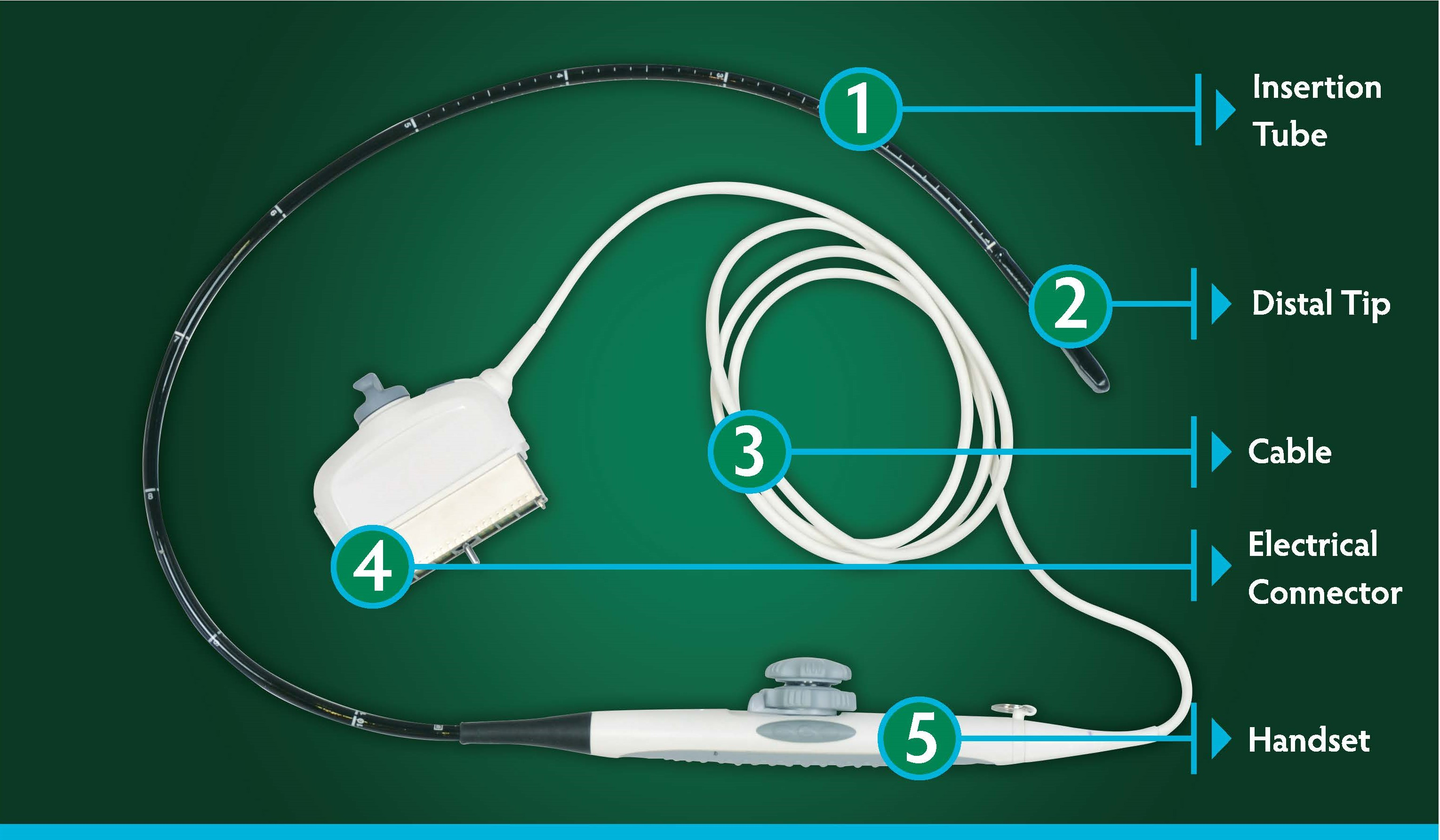
To understand how the procedure works, we must first look at the tool itself. A TEE probe is a marvel of modern biomedical engineering. Let's break down its essential components.
1. The Insertion Tube
The insertion tube is the long, black, flexible portion of the device designed to navigate gently from the mouth into the esophagus. Constructed from durable, medical-grade, biocompatible material, it is highly safe for internal use and easy to disinfect. Along its length, you will find precise centimeter markings. These depth indicators are crucial for the cardiologist, allowing us to position the probe at exact levels of the heart—such as the upper esophagus, mid-esophagus, or the transgastric window within the stomach.
2. The Distal Tip
The distal tip is the technological centerpiece at the "business end" of the probe. Encased within this small tip is a sophisticated array of piezoelectric crystals that power the ultrasound imaging:
They vibrate when an electric current is applied, emitting high-frequency sound waves (ultrasound).
These sound waves bounce off internal structures like heart muscle, valves, and blood cells.
The same crystals then capture the returning echoes and translate them back into electrical signals.
Modern TEE probes utilize multiplane or phased-array transducers, allowing the operator to steer the ultrasound beam electronically through a 180-degree arc without moving the probe itself. Advanced 3D and 4D-capable probes can even reconstruct real-time, three-dimensional models of a beating heart, which is invaluable for pre-surgical planning.
3. The Flexible Cable
Connected to the handle is the robust, flexible cable. It houses a highly shielded bundle of microscopic wires that transmit high-voltage pulses to the transducer crystals at the tip and carry massive amounts of raw echo data back to the processing unit without signal loss or external interference.
4. The System Connector
At the very end of the cable is the electrical connector. This durable, multi-pin plug interfaces directly with the main ultrasound console. It locks securely into the machine, ensuring a high-fidelity connection necessary to process raw signals into high-resolution images in real-time.
5. The Handset (Control Handle)
The handset is the ergonomic steering mechanism held by the cardiologist or anesthesiologist. It features two primary steering wheels:
The Large Knob (Ante/Retroflexion): Controls the tip's forward (nodding) and backward movements to press gently against the esophageal wall.
The Small Knob (Lateral Flexion): Directs the tip left or right.
By combining these steering controls with slight depth adjustments and manual rotations, a skilled operator can safely guide the probe to visualize virtually any cardiac structure.
Section 3: The "Why" - Key Indications for a TEE
A TEE is a specialized problem-solver. It is ordered when clinical questions are too intricate or the structures are too deep for a standard transthoracic echocardiogram (TTE) to capture. Here are the most common scenarios where a TEE is essential.
Indication 1: Detecting Blood Clots (Left Atrial Appendage)
This is a vital pre-procedure test for patients with atrial fibrillation (AFib). Due to the heart's irregular rhythm in AFib, blood can pool and form clots in the Left Atrial Appendage (LAA)—a small, thumb-shaped pouch off the left atrium. If a clot breaks free, it can travel to the brain and cause a stroke.
The Challenge: The LAA sits at the very back of the heart, making it extremely difficult to see clearly via standard chest-wall TTE.
The TEE Solution: Positioned in the esophagus directly behind the left atrium, the TEE probe offers an unobstructed, crystal-clear view of the LAA. We routinely perform a TEE before a cardioversion or ablation to ensure no active clots are present.
Indication 2: Diagnosing Infective Endocarditis (Valve Infections)
When bacteria enter the bloodstream, they can settle on heart valves, creating dangerous clumps of bacteria and tissue debris known as vegetations. This is a life-threatening medical emergency.
The Challenge: These vegetations are often only a few millimeters in size, meaning standard TTE scans can easily miss them.
The TEE Solution: The high-frequency resolution of TEE allows us to detect tiny vegetations early. It also helps us assess secondary complications, such as localized abscesses or valve perforations.
Indication 3: Evaluating Severe Valvular Heart Disease
TEE serves as the gold standard for assessing structural valve damage.
Mitral Valve Repair: Before a surgeon repairs a leaking mitral valve, TEE provides a 3D structural roadmap showing exactly which leaflets are failing.
Prosthetic Valve Dysfunction: If a patient with a mechanical or bioprosthetic valve develops a new murmur or fever, TEE is utilized to check for leaks, clots, or tearing around the valve sewing ring.
Indication 4: Intraoperative Monitoring During Cardiac Surgery
During open-heart surgeries, a TEE probe remains in place throughout the entire operation. This allows the cardiac anesthesiologist to:
Confirm pre-operative findings.
Monitor real-time heart function as the patient is weaned off the cardiopulmonary bypass machine.
Confirm the success of a valve repair immediately before the chest is closed, saving patient lives by catching residual issues early.
Indication 5: Assessing Congenital Defects & Aortic Emergencies
Congenital Defects: TEE provides definitive details on septal defects like ASDs or VSDs, guiding the placement of transcatheter closure devices.
Aortic Dissection: In life-threatening emergencies where the aorta tears, a bedside TEE in the emergency department can yield an immediate, lifesaving diagnosis.
Section 4: The Patient Experience - A Step-by-Step Walkthrough
The idea of "swallowing a tube" understandably causes patient anxiety. However, knowing what to expect can significantly ease your mind. Having guided thousands of patients through this test, here is exactly how the process unfolds.
Step 1: Pre-Procedure Preparation
Fasting (NPO): You must not eat or drink anything for at least 6 to 8 hours before the procedure. Because sedation relaxes your airway and gags reflexes, an empty stomach is critical to prevent vomiting and accidental aspiration into the lungs.
Medications: Discuss your routine medications with your doctor. Most blood pressure and heart medications can be taken with a tiny sip of water, while blood thinners may need to be temporarily paused.
Arrange a Driver: Because you will receive sedatives, you cannot drive yourself home. Plan for a responsible adult to accompany you.
Step 2: In the Procedure Room
You will be monitored closely by a specialized team: the cardiologist, a cardiac sonographer, and a dedicated nurse. The nurse will place an IV line for sedation, EKG patches to track your heart rhythm, a blood pressure cuff, and a pulse oximeter.
Step 3: Performing the TEE Test (15-30 Minutes)
Numbing the Throat: The nurse will spray your throat with a local anesthetic (typically a banana-flavored lidocaine spray) to numb the area and suppress your gag reflex.
Conscious Sedation: You will receive "twilight sedation" through your IV (typically a combination of midazolam and fentanyl). You will remain breathing on your own, feeling deeply relaxed and sleepy, and you likely won't remember the procedure afterward.
Positioning: You will lie comfortably on your left side.
Bite Block: A small plastic ring is placed in your mouth to protect your teeth and safeguard the delicate ultrasound probe.
Inserting the Probe: Once you are fully relaxed, the cardiologist will gently guide the lubricated tip of the probe to the back of your throat and ask you to swallow. This single swallow slides the flexible probe smoothly into your esophagus.
Imaging: You will not feel the probe once it is in place. The lights will dim, and the team will acquire the necessary images over the next 15 to 20 minutes while you rest comfortably.
Step 4: Recovery and Post-Care
The probe is removed in a matter of seconds. You will rest in the recovery area for 30 to 60 minutes. You must not eat or drink until your gag reflex has fully returned to prevent choking. A mild sore throat is the most common side effect and typically clears up within 24 hours.
Section 5: Risks vs. Benefits - A Trustworthy Assessment
No medical procedure is entirely without risk, and being transparent is a key part of healthcare trust. Let's look at the statistics honestly:
Risk Category
Potential Complication
Clinical Likelihood
Prevention & Management
Common & Minor
Sore throat, minor hoarseness, mild temporary gagging
Common (resolves in 24h)
Managed with rest and throat lozenges once numbing wears off.
Uncommon
Minor dental damage, superficial throat scrape, mild aspiration
Rare
Prevented by using the bite block, reporting loose dental work, and strict NPO fasting.
Rare & Serious
Sedation reaction (respiratory depression), esophageal perforation
Extremely Rare (<1 in 5,000)
Prevented by extensive pre-screening for esophageal disease and continuous professional vital monitoring.
In my years of clinical practice, I have never personally encountered a major complication like an esophageal tear. The diagnostic benefits of a TEE—giving us the precise information needed to prevent strokes or plan heart surgeries—overwhelmingly outweigh these minimal, well-managed risks.
Section 6: Conclusion - A Clearer Picture, A Healthier Heart
The transesophageal echocardiogram is a safe, sophisticated, and highly precise imaging tool. By leveraging the natural pathway of the esophagus, TEE bypasses physical barriers to deliver an unparalleled, high-definition look at your heart's inner workings.
While the prospect of the test can seem daunting, the actual experience is highly controlled, quick, and comfortable under twilight sedation. Ultimately, the incredible diagnostic clarity it provides is a critical step toward securing your long-term heart health.
Section 7: Frequently Asked Questions (FAQs)
Q: Will I be completely asleep? Will it hurt? A: You will not be under general anesthesia, but the combination of throat-numbing spray and IV conscious sedation ensures you feel relaxed and pain-free. Most patients fall into a light sleep and have no memory of the probe being inserted.
Q: How long does the entire appointment take? A: Although the imaging itself takes only 15 to 30 minutes, expect to spend about 2 to 3 hours at the clinic or hospital to account for check-in, prep, and post-sedation recovery.
Q: When will I get my results? A: Because the cardiologist performs and interprets the test in real-time, preliminary results can often be shared with you or your driver in the recovery room. A finalized report is sent to your primary care physician or referring doctor within 24 hours.
Q: Can I take a taxi or Uber home alone? A: No. Hospital safety policies strictly prohibit patients from taking rideshares or public transit alone after receiving intravenous sedatives. You must be accompanied by a responsible adult driver who can ensure you get home safely.
Related Articles

Intermittent Ultrasound Faults: Why "Works Fine, Then Doesn't" Is Hard to Fix
Intermittent ultrasound faults are the hardest to diagnose. A field-tested method for reproducing, isolating, and safely returning the system to service.
Why Console Navigation That Gets Worse With Use Often Exposes Panel-Control Weakness Earlier Than A Hard Failure
Console navigation worsening with use? Learn to diagnose panel control board failure and make the right procurement decision—before someone suggests replacing the whole system.
Why A Drifting Keyboard Cluster Usually Points To The Shared Input Path, Not Five Bad Keys
Keyboard keys failing in groups point to a shared input path problem, not bad switches. Learn when a keyboard assembly purchase beats chasing individual keycaps.